published in: Orth Prax 3 (1998) 141 - 146
An anatomically correct component alignment is one of the requirements for a successful total knee arthroplasty. The orientation of the bone resections should follow anatomical landmarks, such as the epicondylar axis, the A/P axis, and the tibial tubercle. This article describes some important anatomical characteristics of the knee joint and their significance for surgical technique.
The knee joint implants that are currently available have reached a high degree of development, and clinical experience has shown that the long-term results of total knee arthroplasty can match those achieved with hip joint replacements.
Aside from the implant design, an accurate surgical technique represents one of the most important factors for a successful knee replacement.
The objectives of total knee arthroplasty include restoration of the natural leg alignment, soft tissue balancing as well as anatomic patella tracking.
It has been indicated in the literature that the majority of implant failures, in particular patella complications and early wear of the polyethylene tibial insert, occur due to surgical error.
This is why it seems beneficial to have a closer look at the anatomical and technical factors which affect correct component alignment.
The following anatomical details play a significant role in understanding the importance of proper implant position:
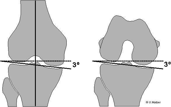
The horizontal plane of a knee joint has a varus slope of approximately 3°, i.e. the tibial joint surface is sloped medially by this amount (6, 9). On the femoral side, this slope results in the medial condyle protruding farther distally (Fig. 1).
 |
| Fig. 1: A/P view of the knee joint surface. |
During knee flexion, the sloped joint line results in internal rotation of the posterior condylar axis by about 4° off the epicondylar axis (so-called "condylar twist" [10]).
The femoral trochlear groove is positioned centrally over the highest point of the intercondylar notch when the epicondylar axis is used for reference; however, in relation to the posterior condyles, the trochlear groove is offset laterally by approximately 3-5 mm (Fig. 2) (13).
The degree of tibial posterior slope shows individual variations; a median value quoted in the literature is about 7 degrees (Fig. 3) (6, 12).
The amount of the Q-angle (Fig. 3) also varies individually; a median value of 15 degrees has been reported (1). In females, the angle is usually somewhat greater.
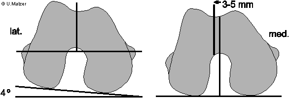 |
| Fig. 2: Position of the patella groove; left: with reference to the epicondylar line; right: with reference to the posterior condyles. 4°: "condylar twist" (see text). |
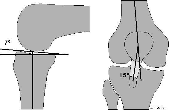 |
| Fig. 3: Left: posterior slope of the tibial plateau. Right: Q-angle (quadriceps angle), which is defined by lines connecting the central point of the patella with the anterosuperior iliac spine above and the center of the tibial tubercle below. |
The anatomical conditions have consequences for bone resections. Basically, there are two options for bone cut orientation (7): In the so-called "anatomical" resection, the same amount of bone is resected from both the medial and the lateral side according to implant thickness (Fig. 4).
The resection follows the anatomical slope of the joint. Therefore, with reference to the mechanical leg axis, the bone cuts are in 3° varus. This is valid for the femur in extension and in flexion, i.e. the bone cuts are always made at the same height.
Since the constant amount of resected bone, which corresponds to the implant thickness, leads to symmetrical gaps, this method usually results in a well-balanced joint in both flexion and extension.
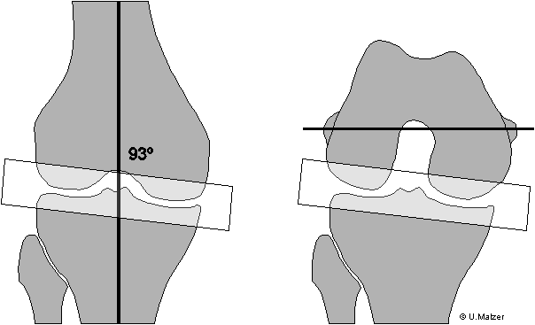 |
| Fig. 4: "Anatomical" resection: The resection line is parallel to the joint line. The amount of resected bone is equal on both sides. The flexion and extension gaps are symmetrical and of a constant width. |
The instrumentation currently available allows for "classical" (or standard) resection (Fig. 5, 6). The distal femur and the proximal tibia are resected perpendicularly to the mechanical leg axis. Usually, this will result in a relative under-resection of the medial tibia and a corresponding over-resection of the medial femur. In extension, these bone cuts are balanced out and generate a symmetrical extension gap.
Observation of the joint line in flexion demonstrates that the posterior femoral cuts have to be asymmetrical because a resection parallel to the posterior condylar axis would result in a trapezoid-shaped flexion gap and a relatively tight joint medially in flexion (Fig. 5). This could be one of the reasons for early postero-medial wear of the polyethylene tibial insert.
To achieve an accurate, asymmetrical resection, a correction of the rotational alignment becomes necessary. This correction corresponds to the slope of the joint and usually amounts to about 3° of external rotation off the posterior condylar axis (Fig. 6).
This correction results once again in a relative medial over- and lateral under-resection of the posterior condyles, which balances out the tibial cut in flexion and generates a symmetrical flexion gap.
An incorrect posterior femoral resection with insufficient external rotation not only results in a tight joint medially, but it also causes the patella groove of the implant to rotate internally, i.e. become medialized (Fig. 7). This increases the Q-angle, which heightens the risk of patella subluxation.
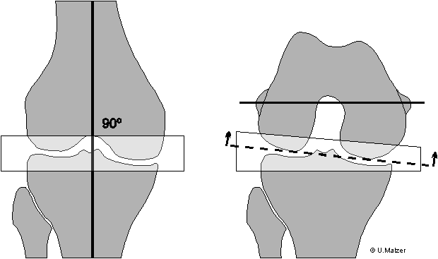 |
| Fig. 5: "Classical" resection without correction of rotation. The distal femoral and the tibial bone cuts are perpendicular to the mechanical leg axis; the posterior femoral cut is parallel to the posterior condylar axis. The result is a trapezoid bone d gap in flexion. |
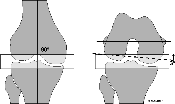 |
| Fig. 6: "Classical" resection with correction of external rotation: a symmetrical flexion gap is established. |
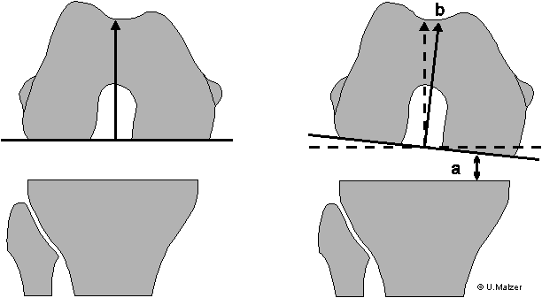 |
| Fig. 7: Left: "Classical resection" with alignment correction. Symmetrical gap, the trochlear groove is positioned centrally over the intercondylar notch. Right: "Classical resection" without alignment correction. Trapezoid-shaped joint gap (a), medialization of the trochlear groove (b). |
Cases with considerable varus or valgus deformities often present bone defects. These are usually located on the medial tibial plateau in the varus knee, and on the lateral femoral condyle in the valgus knee (Fig. 8).
This affects the posterior femoral resection in that the condyles can often not provide a very reliable reference for correct alignment.
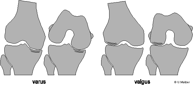 |
| Fig. 8: Bone defects in the varus and valgus knee. |
This is the reason for identifying other anatomic landmarks that can be used for orientation. Two auxiliary lines have proven particularly reliable (Fig. 9):
1. The transepicondylar axis or Insall line (4), which connects the lateral epicondylar prominence and the medial sulcus of the medial epicondyle. The posterior bone cut should be parallel to this line. This resection will usually match that performed with a 3-degree external rotation.
Since the position of the epicondyles remains constant, a posterior femoral resection parallel to the Insall line is not affected by significant bone loss and provides a reliable method even in cases with more severe axial deformities (Fig. 10).
The Insall line has a small disadvantage: it is not always readily identifiable, since the epicondyles are sometimes difficult to palpate under the collateral ligament attachments. Also, it is necessary to determine the true center of the medial epicondyle due to its half-moon shape.
2. As an alternative, the A/P axis or Whiteside line has been defined as the connection of the lowest point of the trochlear groove to the highest point of the intercondylar notch (Fig. 9).
Exact measurements and statistical evaluations of healthy and arthritic femora have shown that the A/P and the epicondylar axis are closely correlated, namely, they are practically always perpendicular to each other.
Since the A/P axis is easier to find, its margin of error is lower than that of the Insall line (3).
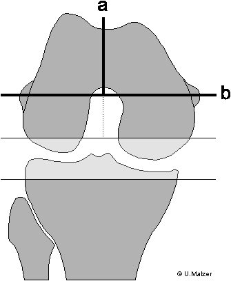 |
| Fig. 9: Epicondylar line (a) and A/P axis (b). |
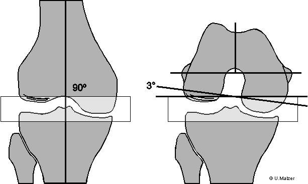 |
| Fig. 10: Correct resection in a valgus knee. A 3-degree correction would not be sufficient to create a symmetrical flexion gap. |
Two factors are of particular importance for tibial component alignment:
The posterior slope contributes to the overall flexion gap width. An insufficient posterior slope will cause a relative tightness and a flexion deficit, while a slope which is too steep will result in instability in flexion and a more pronounced roll-back of the femoral component.
This might lead to overloading of the posterior tibial insert along with increased polyethylene wear (11). It is recommended to select an angle, which will be smaller than the anatomical angle of about 7 degrees (12). It seems reasonable not to exceed a posterior slope of about 3-5 degrees.
To establish the posterior slope, orientation to the anterior crest of the tibia rather than the joint surface is somewhat more exact because of the longer reference distance. Mathematically, moving the alignment line at the joint surface by 1 mm [0.04"] generates an angle of 1.2 degrees, while a difference of 1 cm [0.4"] over the entire length of the tibia only results in a deviation of 1.5 degrees (Fig. 11).
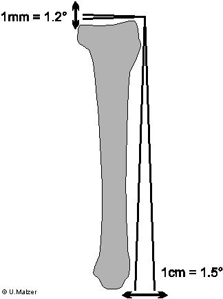 |
| Fig. 11: The anterior crest of the tibia provides a reliable reference for establishing the posterior slope of the tibial plateau. |
The rotational alignment of the tibial component also contributes to proper patella tracking. An internal rotation of the component must be avoided as it will lead to external rotation of the tibial tubercle, increase the Q-angle, and result in lateralization of the patella (Fig. 12).
There are several landmarks to aid orientation (fig. 13). The most reliable site is the medial border or medial third of the tibial tubercle (8). It is easy to find and has direct correlation with patella tracking.
The often cited orientation to the center of the talus or the third metatarsal includes the risk of internal rotational misalignment: Particularly when a foot holder is used, the foot is held in position anteriorly, while the patella ligament externally rotates the head of the tibia when the patella is laterally subluxated. This might lead to incorrect positioning of the tibial component in internal rotation (Fig. 14).
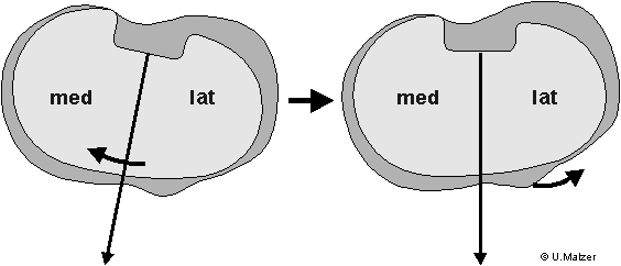 |
| Fig. 12: Internal rotation of the tibial component (left) leads to external rotation of the tibial tubercle (right). |
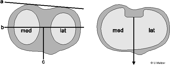 |
| Fig. 13: Reference lines for the rotational alignment of the tibial component. a: posterior condylar axis; b: transcondylar line; c: tubercle line. Right: Referencing the tubercle line provides the most reliable method of tibial component alignment. |
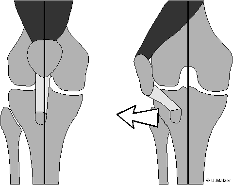 |
| Fig. 14: Intraoperative lateral subluxation of the patella results in external rotation of the head of the tibia. |
Patella tracking remains one of the toughest challenges of total knee arthroplasty. Approximately 30 to 40 percent of all complications occur at the patello-femoral articulation. The discussion above highlights the importance of proper femoral and tibial alignment, which is a prerequisite for anatomic patella tracking.
When implanting a patella, it is also very important to slightly medialize the component, as this position replicates the natural position of the apex of the patella (14). Small implants are recommended for small-sized patellas as this will allow for adequate medialization of the component.
In addition, patella tracking can be improved in individual cases by offsetting the femoral component laterally by a few millimeters (Fig. 15).
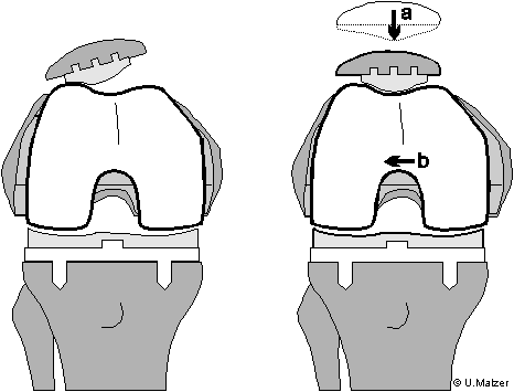 |
| Fig. 15: Improved position of the patella through medialization (a) and a slight lateralization of the femoral component. |
Total joint arthroplasty designs have improved significantly over the past few years, and modern instrumentation allows for very accurate implantation. One should not, though, rely exclusively on the instruments.
Establishing proper component alignment by referencing anatomical landmarks is vitally important for a successful operation, particularly in cases with severe deformities, bone defects, and ligamentous instability.
It is only after the appropriate bone cuts have been made that ligamentous stability should be examined and, if necessary, soft tissue balancing (ligament releases etc.) performed.
By taking the guidelines described above into consideration, we were able to see improved clinical results in our patients. It has become exceedingly rare to perform a release of the lateral capsule or tibial tubercle osteotomy. Correct implant position and adequate soft tissue balancing also facilitate patient postoperative care and mobilization.
1. Aglietti, P., Insall, J.N., Cerulli, G.: Patellar pain and incongruence. I:Mesurements of incongruence. Clin. Orthop. 176(1983) 217-224
2. Anouchi, Y.S., Whiteside, L.A., Kaiser, A.D., Milliano, M.T.: The effects of axial rotational alignment of the femoral component on knee stability and patellar tracking in total knee arthroplasty demonstrated on autopsy specimens. Clin.Orthop. 287 (1993) 170-177
3. Arima, J., Whiteside, L.A., McCarthy, D.S., White, S. E.: Femoral rotational alignment, based on the anteroposterior axis, in total knee arthroplasty in a valgus knee. J. Bone Joint Surg. (A) 77,9 (1995) 1331-1334
4. Berger, R.A., Rubash, H.E., Seel, M.J., Thompson, W.H., Crossett, L.S.: Determining the rotational alignment of the femoral component in total knee arthroplasty using the epicondylar axis. Clin. Orthop. 286 (1993) 40-47
5. Hsu, R.W., Himeno, S., Coventry M.B., Chao, E.Y.: Normal axial alignment of the lower extremity and load-bearing distribution at the knee. Clin. Orthop. 255 (1990) 215-27
6. Krackow, K.A. : Approach to planning lower extremity alignment for the total knee arthroplasty and osteotomy about the knee. Adv. Orthop. Surg. (1983) 69- 88
7. Krackow, K.A.: The technique of total knee arthroplasty. Mosby, St.Louis, 1990
8. Lotke, P.A.: Knee Arthroplasty. 6:Standard principles and techniques. In: Thompson, R.C. (ed.):Master techniques in orthopaedic surgery. Raven Press New York 1995
9. Moreland, J.R., Basset, L.W., Hanker, G.J.: Radiographic analysis of the axial alignment of the lower extremity. J. Bone Joint Surg. (A) 69,5 (1987) 745-749
10. Ranawat, C.S., Rodriguez, J.A.: Malalignment and malrotation in total knee arthroplasty. In: Insall, J.N., Scott, W.N., Scuderi, G.R. : Corrent concepts in primary and revision total knee arthroplasty. Lippincott-Raven, Philadelphia 1996
11. Wasielewski, R.C., Galante, J.O., Leightly, R.M., Natarjan, R.N., Rosenberg, A.G.: Wear patterns on retrieved polyethylene tibial inserts and their relationship to technical considerations during total knee arthroplasty. Clin. Orthop. 299 (1994) 31-43
12. Whiteside, L.A., Amador, D.D.: The effect of posterior tibial slope on knee stability after Ortholoc total knee arthroplasty. J. Arthroplasty 3 (1988) Suppl:51- 57
13. Whiteside, L.A., Arima, J.: The anteroposterior axis for femoral rotational alignment in valgus total knee arthroplasty. Clin.Orthop. 321 (1995) 168-172
14. Yoshii, I., Whiteside, L.A., Anouchi, Y.S.: The effect of patellar button placement and femoral component design on patellar tracking in total knee arthroplasty. Clin. Orthop. 275 (1992) 211-219